|
BOOK NOW |
ASK ABOUT YOUR PAIN |
Home > Blog > Physiotherapy > Conditions > Shoulder Pain > Shoulder Blade Pain > Proximal Humerus Fracture
Proximal Humerus Fracture

Proximal humeral (humerus) fractures happens when the top aspect of the upper arm bone, that is shaped like a ball, is fractured or have a break.
It's also commonly called a shoulder fracture, which are categorized and grouped according to
- location of break on the humeral bone
- how many pieces it broke into (2, 3, 4 or more pieces)
- if the shoulder humeral fracture includes movement (translation and/or dislocation).
Typically, the most common cause of proximal humerus fractures are falling onto an outstretched hand as well as being involved in road traffic or motor vehicle accidents which includes high force or speed.
Good news is that most of the time, proximal humeral fractures does NOT require any corrective surgical operations and usually treated with immobilization and rest first in a sling, followed by shoulder fracture rehabilitation for a period of time to improve shoulder movement and strength.
humerus bone review

Our humerus / humeral bone refers to the long bone in our upper arm that sits between our shoulder and elbow, and the very top part of it is called the proximal humerus.
Proximal is a medical and anatomical term meaning "nearer to midline" - so in this case, proximal humerus means that it's the part of humerus that is closest to our midline, and that is essentially the humeral head.
Next, we have four (4) rotator cuff muscles which attach to the humerus head, and they're the main muscles involved and responsible for shoulder movement, stability and strength. Whenever our rotator cuff muscles or tendons are injured, it leads to lots of inconveniences and pain.
Here are the most important components to our proximal humeral bone:
Humeral Head
The top of the humerus is shaped pretty much like a ball (although to me it's a modified ball, it looks very similar to if you make a fist with you hand and then flex your wrist - that's very similar to how it looks and feels) and it is known as the head of humerus or humeral head.
Our humeral head rests in a fairly shallow socket that is formed by part of
the shoulder blade, known as the glenoid fossa to form the glenohumeral
joint which is our shoulder joint.
Greater Tubercle/Tuberosity
Our greater tubercle refers to a large nodule (pure mass of bone) found on the outward facing side of the proximal humerus. Three of the four rotator cuff attach to the greater tubercle
- supraspinatus
- infraspinatus and
- teres minor
Lesser Tubercle/Tuberosity
The lesser tubercle of the humerus refers to a smaller nodule, and this time it is found on the front side of the proximal humerus. The fourth rotator cuff muscle called the subscapularis, attaches here.
Anatomical Neck
The anatomical neck of the humerus is a diagonal groove that lies and is located between the head of humerus and the greater and lesser tubercles. The articular capsule of the shoulder joint attaches here.
Surgical Neck
The surgical neck of our humeral bone runs horizontally (straight across), just below the greater and lesser tubercles at the bottom of the humeral head. Most proximal humerus fractures typically happen at the surgical neck.
what are the typical Causes of Proximal Humerus Fractures?
Proximal humerus fractures account for a signifiant large majority of fractures of the upper arm and is actually 5% of all fractures.
Most common causes of a proximal humerus fracture include:
A Fall / Falls
As mentioned earlier, the most common cause of a proximal humerus fracture is falling over onto an outstretched hand (this is often abbreviated to "FOOSH").
In fact, what patients need to know is that the force or fall doesn’t need to be from any great
height; "just" falling from a standing height can be enough to break the proximal humeral bone.
Of course, patients falling down stairs is also a common cause of the same fracture (and others)

A Direct Blow
A high-energy blow to the shoulder, such as a
- sports injuries such as a sporting tackle or collision or
- road traffic accident / motor vehicle accidents
can also cause a proximal humerus fracture. Often in these high speed or power circumstances our shoulder joint can also dislocate, and what this means is that the head of humerus slips out of the glenoid socket.
(Older) Age
Approximately 75% of proximal humerus fractures occur in people over the age of 60 because our bones will get weaker and more brittle with age, making them more just easier to break and fracture.
Fractures of the proximal humerus are the third most common type of broken bone in the 65’s after hip fractures and wrist fractures.
In osteoporosis the bone becomes increasingly brittle and weakens, making it more likely to break. A milder version of this is low bone mineral density (osteopenia) which also increases fracture risks.
Gender
Women are three (3) times more likely to experience a proximal humeral fracture than men - this could be due to variable factors such as:
- muscle strength and size to absorb force
- higher risk of osteoporosis or osteopenia
- balance issues
- etc
Classification of Humerus Fractures
A proximal humerus fracture is typically categorised using the Neer System which takes into considerations
- the number of fracture parts and
- the level of “displacement” of these fractured humeral parts
Neer divides the proximal humerus into four parts:
- the anatomical neck
- the greater tuberosity
- the lesser tuberosity and
- the surgical neck
Neer then looks at the level of displacement of the parts: if there is more than 1 cm of separation or the angulation is greater than 45 degrees of a part, it is considered a displaced fracture.
One-Part Fractures
A one-part proximal humerus fracture means that there is no displacement.
Any one or more of the four parts may be fractured, but there is less than 1 cm separation between the two fractured ends and less than 45 degree angulation.
For example there could be a fracture of the surgical neck and the
greater tuberosity, but as long as neither of the parts are displaced,
it is still classed as a one part fracture.
Fortunately most (up to 75%) of proximal humerus fractures are one part fractures
Two part fractures are where one part is displaced more than 1 cm or rotated more than 45 degrees. Again, there may be multiple fractures in multiple parts, but we're referring to at least one part is displaced here.
Approximately 20% of proximal humerus fractures are two part fractures.
Three Part Fractures
Three part fractures are where two parts are displaced.
There will most likely be displacement of the surgical neck and one of the tuberosities (most of the time it involves the greater tubercle), while the other humeral tuberosity remains attached.
Approximately 5% are three part fractures.
Four Part Fractures
This is where all four parts are fractured with three of them displaced in relation to the fourth.
Fortunately, four part fractures are very, very rare (less than 1% chance) but when it happens, it is classified as very severe injury and almost always will require corrective shoulder surgery - patients are highly recommended for surgery unless medically not able to.
Fracture Dislocation
Fracture dislocations happen when the shoulder is dislocated with a proximal humeral fracture, meaning it has come out of its socket, as well as fractured shoulder.
A proximal humerus fracture dislocation may be a two, three or four part fracture.
Symptoms Of A Proximal Humerus Fracture
Proximal humerus fractures don’t just happen without any forms of external forces, and most patients will be able to recall and connect the specific bone fracture to a specific event, date and place.
A proximal humerus fracture will typically cause:
- Pain: The pain from the shoulder injury and bone fracture will be near instant, severe and ongoing. Patients will know for sure 100% that it's definitely injured and possibly fractured.
- Limited Shoulder And Arm Movement: Patients will experience pain with any upper arm and shoulder movement (will be extremely painful) so movement will be very restricted
- Swelling And Bruising: There will be significant swelling and bruising in the upper arm which may extend all the way down to the hand within a couple of days of injury
- Bony Sounds (Crepitus):
Patients may be alarmed when they hear and even experience grinding or grating noise or sensation when moving the ir broken humeral
arm. The reason for these sounds and sensations is caused by the broken pieces of bone fragments that are rubbing against each
other when moved
- Obvious Fracture Deformity: If
the patient's shoulder fracture includes is a displaced fracture, the arm may appear deformed or just “not
looking quite normal or right”. If there is a fracture dislocation, the deformity will be
obviously visible even to lay person
- Bleeding: If it unfortunately involves the fractured part piercing and puncturing the skin (termed as an open fracture) there will be bleeding
- Altered Sensation: In some situations, the sharp bony fragments may injure the nerves around the shoulder and that may result in altered sensation such as pins and needles or numbness below the fracture site. Less than half of all proximal humerus fractures have associated nerve damage but it tends to be the axillary nerve that is most often affected if any.
Diagnosing Humeral Fractures

If our senior physiotherapists or your shoulder doctor suspects a proximal humerus fracture, you will definitely be referred and sent for at least one (1) types of imaging scans to evaluate the severity of the fracture, where exactly it is and how many parts and if there is any dislocation or movement of the fractured parts.
Typically, taking x-rays with different
angles of the shoulder and upper arm are enough to diagnose and
categorize a humerus fracture.
X-rays are usually taken in three perpendicular planes:
- the front (true AP view)
- the side (lateral
scapula Y view) and
- through the armpit (axillary view)
In some cases, you may also be sent for a CT or MRI scan if the x-rays imaging results aren’t clear or aren't clear enough. If the greater tuberosity is fractured then an ultrasound scan will be performed to check for rotator cuff muscle and/or tendon damage.
Treatment of A Proximal Humerus Fracture
Conservative / Non-Operative Approach
Good news is that in most cases, a proximal humerus fracture will NOT require surgery. Nearly 80% of proximal humeral fractures are undisplaced or only minimal displaced fractures and therefore non-operative treatment is appropriate. Non-operative treatment for a proximal humerus fracture consists of:
1) Immobilisation
For the first few weeks after a proximal humerus fracture, patients are required to keep their affected arm in a sling that is worn to hold the fracture still and to allow the swelling and pain to subside.
The shoulder fracture needs to be completely immobilised for at least 10-14 days to allow time for the bones to start slowly joining back together and heal, so the sling must be worn all the time.
Yes, we mean day and night.
After this, you can gradually reduce how much you wear the sling and by around 12 weeks you shouldn’t need it at all (yay!).
Most of the time, patients are prescribed a shoulder immobiliser or sling and swath. These are good such that because they provide support to the elbow and forearm and counteract the weight of the arm. The extra strap around the body helps to keep the upper arm immobilised.
In some cases, patients may instead be issued a collar and cuff instead. These provide less
support but allow the weight of the arm to provide gentle traction which
can help improve the alignment of the fracture.
2) Medication
Medication to relieve pain and inflammation will be prescribed by your doctor such as NSAIDS – non-steroidal anti-inflammatories e.g.
- ibuprofen or
- naproxen
If you have an open fracture or surgery you will also be given antibiotics to reduce the risk of infection.
3) Exercises
For the first couple of weeks after a proximal humerus fracture the shoulder should be immobilised in the sling, but please note that it is very, very important to do your
- elbow
- wrist
- finger
exercises to prevent any stiffness or weakness.
It's extra painful to have stiffness in shoulder post shoulder fracture AND stiffness in elbows, wrists and fingers...
Typically you're allowed to take your arm out of the sling to do these exercises.
These exercises should
be started as soon as possible and be done at least 3 - 5 times a day, if not more. It
is important to move the elbow, wrist and fingers through their full
range of motion to prevent stiffness and hand grips and balls are a
great way to maintain strength.
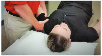
After about two (2) weeks, you should be able to start gentle exercises to get the shoulder moving and our senior physiotherapists will work on a shoulder physiotherapy program together with you.
Pendulum exercises are the best place to start for regaining mobility. These are exercises where you use gravity to move the arm so your muscles don’t have to do anything, so as to ensure that your muscles they don’t catch onto or pull on the fracture site.
Then after around three weeks you can start active assisted exercises (AAROM) where you use your good arm or a bar/stick to support and move the broken arm. You may also be prescribed a shoulder pulley system to use.
In terms strengthening, you will start with isometric exercises.
These are exercises where you push against resistance but without
any arm or muscle movement and it's purposely planned this way so you don’t aggravate the fracture site. You may be
allowed to start these as soon as pain allows or you may need to wait
until the fracture has fully healed – you will have to check with your doctor or with our senior physiotherapists.
After about 6 - 10 weeks the fracture parts should have united and you can move on to active mobility exercises and progressive strengthening exercises which may include resistance band exercises to regain rotator cuff strength.
Shoulder physiotherapy will continue until you have regained full
- movement
- strength and
- stability
in the shoulder and elbow.
You may need to keep doing exercises for up to a year (yes up to 12 months) to achieve this. Do note that unfortunately not everyone does regain full range of movement after a proximal humerus fracture and some people will have ongoing difficulty lifting their arm above head height (but we will always aim for 100% recovery of course).
It is very very important to rigorously follow your shoulder physiotherapy exercise program and do your shoulder exercises daily to ensure you regain as much
- range of motion
- flexibility and
- strength as possible
Failure to do so will lead to reduced shoulder movement which will likely cause pain, discomfort and ultimately will affect function.
Your shoulder doctor will see you regularly and x-ray the shoulder periodically to check on the healing process. Everyone heals at different rates so follow the guidance of your doctor or our senior physical therapist as to when you can progress to the next stage of rehab.
Surgical Approach
Unfortunately around 15% of proximal humerus fractures tend to be displaced and require surgery.
In most cases, metalwork is used to realign and fix the bones. This is a temporary measure to hold things together to allow the bone time to heal, and these are not meant to be a permanent support structure. The metal work may be removed at a later date if it comes loose or causes any problems. If the bone fails to unite, then further surgery will be required.
There are a few different types of surgery for a proximal humerus fracture depending on the type of fracture and the individual’s functional needs.
Closed Reduction
A closed reduction is where a proximal humerus fracture is realigned under anaesthesia without having to open up the shoulder with a scalpel (typically termed as MUA = manipulation under anesthesia).
This may be all that is needed, but if the MUA realignment proves to be unstable, then the surgeon may use percutaneous pins or K-wires to hold the bone in position. These are inserted through the skin and into the bone at various angles to hold the fracture still.
In some cases, the ends of the pins or K-wires are left slightly exposed so they can easily be removed once callus formation has begun, however there are increased risks of infection and pin movement (termed as migration or displacement), so buried pins may be safer to be used instead.
A closed reduction is most commonly done for two-part surgical neck fractures
ORIF (Open Reduction Internal Fixator)
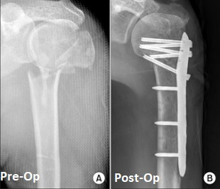
ORIF is the abbreviation for open reduction internal fixation and is where the fracture is stabilized and fixed using a metal plate and screws.
The orthopedic surgeon will make an incision at the front or side of your shoulder. If there is any fracture displacement, then the fracture will be reduced (brought together and realigned).
A metal plate is then inserted and held in place by surgical screws above and below the fracture site to keep it stable and secure. After that, the incision is then closed with staples or sutures (typically with sutures).
An ORIF is most commonly done on younger patients for two, three or four part fractures or when there is displacement of the greater tuberosity.
Intramedullary (IM) Nailing

A small anterolateral (front and side) incision is made through the skin and through all underlying soft tissues.
The orthopedist will then insert a rod-shaped nail into the hollow centre of the humerus bone, known as the medullary canal and held in place with surgical screws. This splints the fracture from the inside to hold it steady.
Intramedullary nails are typically selected when / for a two part or three part proximal humerus fracture, or if there is also damage to the humeral shaft below.
Shoulder Hemiarthroplasty

A shoulder hemiarthroplasty (also means partial shoulder replacement), involves removing the humeral head and replacing it with a metal implant.
Then the stem of the implant is inserted into the medullary canal (the hollow middle of the upper arm bone) and will either be wedged tightly in place or secured with special bone glue cement.
The shoulder head "ball" is then attached to the top of the stem.
Most of the time, a shoulder hemiarthroplasty is done where the fracture is especially complex or if other treatment methods have failed.
Maybe it is an anatomical neck fracture, a four-part fracture, a fracture dislocation, or the rotator cuff may have also been damaged.
Also, if the blood supply to the humeral head has been
compromised plus if there is a high risk of osteonecrosis (where the bone starts to rot and dies), then your shoulder surgeon will likely refer a hemiarthroplasty to be the treatment of choice.
Total Shoulder Arthroplasty

A total shoulder arthroplasty refers to where both parts of the shoulder joint are replaced (the arm and the body part where the arm attaches to part).
The ball-shaped humeral head is removed and replaced with a metal implant. The implant is shaped like a half moon and has a stem which runs down the middle of the humerus bone.
The socket part of the joint on the shoulder blade is shaved down and a plastic socket is positioned and held in place with special cement.
Shoulder replacement is not always the first option by the orthopedic surgeon - it will be done only if there is BOTH damage to
- the shoulder blade
socket (the glenoid) together with
- the proximal humerus fracture
The glenoid fossa / process may have been damaged at the time of injury or there may be significant arthritis (wear-and-tear, degeneration) in the joint that would make a partial shoulder replacement unsuitable.
Our rotator cuff muscles and tendons must be intact for a total shoulder arthroplasty, if not there is a high risk of loosening of the prosthesis.
93% of total shoulder replacements typically last at least 10 years.
Reverse Shoulder Replacement
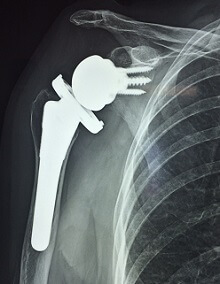
If the patient's rotator cuff tendons and/or has been damaged (partially torn or completely ruptured) with the proximal humerus fracture, then the shoulder surgeon may recommend a reverse shoulder replacement.
It is most suitable for people over the age of 70 who have low shoulder movement and strength needs.
One requirement is that the deltoid muscle of the shoulder must be functioning well, strong and in good working order.
In a reverse shoulder replacement,
- the ball part of the implant is placed on the shoulder
blade and
- the socket is placed on the humerus - this is the opposite way round to normal
(that's why it's called "reverse")
That may sound like a strange thing to do, but what it does is allow the deltoid muscle to make up for the deficiency in the rotator cuff to allow greater shoulder movement than would otherwise be possible.
Recovering From A Proximal Humerus Fracture
Typically, the rehabilitation and recovery process following a proximal humerus
fracture is basically the same even if patient was treated with or without surgery.
The most important part of the recovery process is improving and regaining their shoulder range of
movement.
Phase 1: Weeks 0 - 4
During this phase, patients will be have their affected shoulder and arm immobilized and protected in a sling, which needs to be worn all the time (yes, night, day and sleeping time) except when
- showering
- dressing change
- doing exercises
Patients will be taught a few exercises:
- shoulder pendulum exercises
- active range of motion exercises for unaffected joints (elbow, wrist, fingers etc)
Phase 2: Weeks 4 - 10
Patient will continue to review with their treating shoulder surgeon and x-rays will be done regularly, once the fracture has started to unite with no dislocation or displacement, then we can up the exercises:
- actively-assisted shoulder exercises, and the amount of assistance and support is progressively weaned off
- patient is allowed shoulder involvement for gentle functional movement and use, without pain
Phase 3: Week 10 Onwards
At this stage, patient can progress to more "strong" strengthening exercises. We will often start with isometric strengthening and gradually progress to strengthening exercises with shoulder/arm movement. This will continue to progress until patient has achieved 100% recovery in shoulder
- strength
- mobility / movement
As a general guideline patients can expect:
- Week 1: to be able to write and type, use phone etc
- Week 6 - 12: for the bone to have fully healed and you should no longer need to use your sling
- Week 12 (3rd Month): to be able to reach up to get things from a cupboard and hang the washing on the line
- Week 12 - 24 (3 - 6 months): to get back to lifting without any restriction
- Week 26 to 52 (6 - 12 months): to have regained full function if the arm – shoulder mobility might always be partly restricted

On smoking:
Please note that research has shown that smoking (or using nicotine products) directly delays bone healing and can increase the risk of non-union by around 6 times (this is 600% increase of bone fracture issues).
You see, what happens is that nicotine restricts the size of blood vessels by around 25% which means the bone gets less of the oxygen and nutrients it needs to heal, which can significant slowdown the healing process.
what are potential Complications related to Proximal Humerus Fractures
The most common complications that a patient may experienced with a proximal humerus fracture injury includes:
Decreased Shoulder Movement And Function
Full strength and mobility may never be achieved after a proximal humerus fracture, particularly in elderly patients or those who do not follow their shoulder physiotherapy exercise program.
Patients may take up to 12 months (1 year) to regain their full shoulder movement and strength with shoulder physiotherapy
Also known as adhesive capsulitis, frozen shoulder happens when our shoulder joint capsule thickens and tightens which can cause:
- shoulder pain
- very limited shoulder movement
Most of the time, this can happen when the shoulder
is not moved (immobilised or protected) for a long period of time. Starting a shoulder physiotherapy exercise program as
early as possible helps to reduce the risk of developing a frozen
shoulder.
Avascular Necrosis (AVN)
A proximal humerus fracture can disturb the blood supply to the humeral head resulting in avascular necrosis aka osteonecrosis. What happens is that because of the impaired blood flow and supply to and from the humeral head, the bone cells gradually rot, wither and die.
The humeral head gradually collapses resulting in pain and reduced movement.
Blood
flow to the humeral head comes from vessels that branch off the axillary
artery and travel upwards from the armpit region. Anatomical neck
fractures and four part fractures are the most likely to impede blood
flow and cause avascular necrosis.
Damage to the shoulder joint increased the risk of developing early shoulder osteoarthritis, which can be very painful and affects daily function.
Surgical Risks
Any surgery has the risk of
- infection
- nerve damage
- poor bone
healing (eg non union, mal union)
- wound healing problems
- loosening of the prosthesis
Reference Sources: