|
BOOK NOW |
ASK ABOUT YOUR PAIN |
Home > Blog > Spinal Physiotherapy > Degenerative Disc Disease Physiotherapy
Degenerative Disc Disease Physiotherapy

Degenerative disc disease (DDD) is a medical condition (ICD-10-CM M51.35-37) in which there are anatomic changes and a loss of function of varying degrees of one or more intervertebral discs of the spine of sufficient magnitude as to cause symptoms.
The root cause is thought to be loss of soluble proteins within the fluid contained in the disc with resultant reduction of the oncotic pressure, which in turn causes loss of fluid volume. Normal downward forces cause the affected disc to lose height, and the distance between vertebrae is reduced.
The anulus fibrosus, the rigid outer shell of a disc, also weakens.
This loss of height causes laxity of the longitudinal ligaments, which may allow anterior, posterior, or lateral shifting of the vertebral bodies, causing
- facet joint malalignment and arthritis
- scoliosis
- cervical hyperlordosis
- thoracic hyperkyphosis
- lumbar hyperlordosis
- narrowing of the space available for the spinal tract within the vertebra (spinal stenosis)
- narrowing of the space through which a spinal nerve exits (vertebral foramen stenosis) with resultant inflammation and impingement of a spinal nerve, causing a radiculopathy.
DDD can cause mild to severe pain, either acute or chronic, near the involved disc, as well as neuropathic pain if an adjacent spinal nerve root is involved. Diagnosis is suspected when typical symptoms and physical findings are present; and confirmed by x-rays of the vertebral column.
Occasionally the radiologic diagnosis of disc degeneration is made incidentally when a cervical x-ray, chest x-ray, or abdominal x-ray is taken for other reasons, and the abnormalities of the vertebral column are recognized.
The diagnosis of DDD is not a radiologic diagnosis, since the interpreting radiologist is not aware whether there are symptoms present or not. Typical radiographic findings include disc space narrowing, displacement of vertebral bodies, fusion of adjacent vertebral bodies, and development of bone in adjacent soft tissue (osteophyte formation). An MRI is typically reserved for those with symptoms, signs, and x-ray findings suggesting the need for surgical intervention.
Treatment may include spinal physiotherapy to
- reduce pain and increase any reduced range of motion of the spine
- strength training with emphasis on correcting abnormal posture, assisting the paravertebral (paraspinous) muscles in stabilizing the spine
- core muscle strengthening
- stretching exercises
- massage therapy
- oral analgesia with non-steroidal anti-inflammatory agents (NSAIDS)
- topical analgesia with lidocaine
- cold therapy
- heat therapy
- ultrasound therapy
- computerized spinal decompression therapy
Immediate surgery may be indicated if the symptoms are severe or sudden in onset, or there is a sudden worsening of symptoms. Elective surgery may be indicated after six months of conservative therapy with unsatisfactory relief of symptoms.
Signs and symptoms
Degenerative disc disease can result in lower back pain or upper neck pain.
In fact, the amount of degeneration does not correlate well with the amount of pain patients experience. Many people experience no pain while others, with exactly the same amount of damage have severe, chronic pain.
Whether a patient experiences pain or not largely depends on the location of the affected disc and the amount of pressure that is being put on the spinal column and surrounding nerve roots.
Nevertheless, degenerative disc disease is one of the most common sources of back pain and affects approximately 30 million people every year. With symptomatic degenerative disc disease, the pain can vary depending on the location of the affected disc. A degenerated disc in the lower back can result in lower back pain, sometimes radiating to the hips, as well as pain in the buttocks, thighs or legs. If pressure is being placed on the nerves by exposed nucleus pulposus, sporadic tingling or weakness through the knees and legs can also occur.
A degenerated disc in the upper neck will often result in pain to the neck, arm, shoulders and hands; tingling in the fingers may also be evident if nerve impingement is occurring.
Pain is most commonly felt or worsened by movements such as sitting, bending, lifting and twisting.
After an injury, some spinal discs become painful because of inflammation and the pain comes and goes. Some people have nerve endings that penetrate more deeply into the anulus fibrosus (outer layer of the disc) than others, making discs more likely to generate pain.
In the alternative, the healing of trauma to the outer anulus fibrosus may result in the innervation of the scar tissue and pain impulses from the disc, as these nerves become inflamed by nucleus pulposus material.
Degenerative disc disease can lead to a chronic debilitating condition and can have a serious negative impact on a person's quality of life. When pain from degenerative disc disease is severe, traditional nonoperative treatment may be ineffective.
Cause
There is a disc between each of the vertebrae in the spine. A healthy, well-hydrated disc will contain a great deal of water in its center, known as the nucleus pulposus, which provides cushioning and flexibility for the spine.
Much of the mechanical stress that is caused by everyday movements is transferred to the discs within the spine and the water content within them allows them to effectively absorb the shock. At birth, a typical human nucleus pulposus will contain about 80% water.
However natural daily stresses and minor injuries can cause these discs to gradually lose water as the anulus fibrosus, or the rigid outer shell of a disc, weakens.
This water loss makes the discs less flexible and results in the gradual collapse and narrowing of the gap in the spinal column. As the space between vertebrae gets smaller, extra pressure can be placed on the discs causing tiny cracks or tears to appear in the anulus. If enough pressure is exerted, it's possible for the nucleus pulposus material to seep out through the tears in the anulus and can cause what is known as a herniated disc.
As the two vertebrae above and below the affected disc begin to collapse upon each other, the facet joints at the back of the spine are forced to shift which can affect their function.
Additionally, the body can react to the closing gap between vertebrae by creating bone spurs around the disc space in an attempt to stop excess motion. This can cause issues if the bone spurs start to grow into the spinal canal and put pressure on the spinal cord and surrounding nerve roots as it can cause pain and affect nerve function. This condition is called spinal stenosis.
For women, there is good evidence that menopause and related estrogen-loss are associated with lumbar disc degeneration, usually occurring during the first 15 years of the climacteric. The potential role of sex hormones in the etiology of degenerative skeletal disorders is being discussed for both genders.
Degenerative Disc Disease Physiotherapy
Following diagnosis, the senior physiotherapist will then create a custom physio intervention plan for you, and depending on the causes of the problem, you may be treated with a combination
- heat therapy to loosen the neck muscles, tissues and joints
- cold therapy to decrease inflammation and pain
- ultrasound therapy to accelerate soft tissue healing
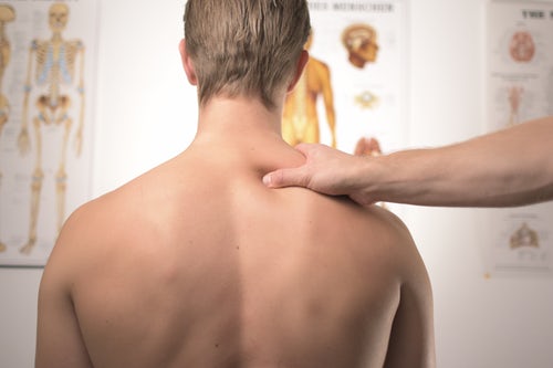
- manual therapy
- joint manipulation and mobilization to increase the natural range
- pain relief physiotherapy
- stretching and flexibility physiotherapy
- dry needling physiotherapy
- computerized decompressive spinal traction
- neck strengthening
- core stability strengthening